India Unveils Detailed 3‑D Atlas of the Human Brainstem
For more than a century, neuroscientists have mapped the human brain much as early cartographers mapped unknown lands, piecing together a vast landscape from scattered observations. The challenge remains: a brain weighing 1.2‑1.5 kg contains 86 billion neurons, but pathologists can only sample a handful of tissues during diagnosis.
The Sudha Gopalakrishnan Brain Centre (SGBC) at IIT Madras has taken a crucial step towards filling this knowledge gap by creating the world’s most detailed cellular‑resolution 3‑D map of the brainstem. Launching the Atlas of Neurochemical Characterisation of the Human Brainstem with 3D Reconstruction—shortened to Anchor—the team combined more than 500 tissue sections from foetal, childhood and adult specimens into a single digital atlas. The reconstructed volume highlights over 200 clusters of neurons and nerve pathways, using eight chemical markers to distinguish cell types.
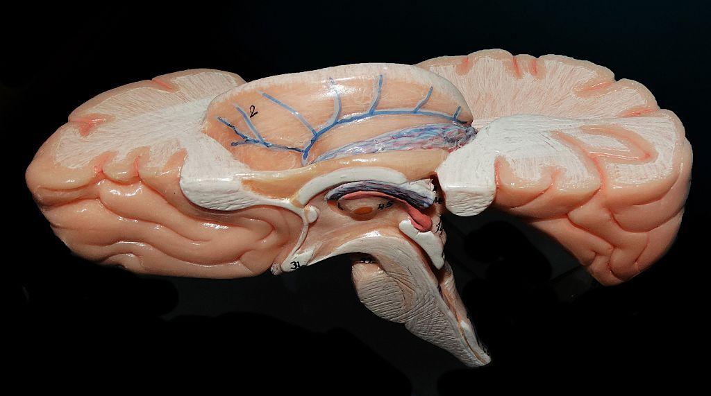
The brainstem, though only a sliver of the brain, is essential for life, connecting the brain to the spinal cord and regulating breathing, heartbeat, sleep, wakefulness and movement. Its densely packed architecture has made exhaustive mapping difficult until now.
Anchor’s importance lies in linking two traditionally separate worlds: whole‑brain MRI imaging and cellular pathology. "We are seeing a visionary programme that puts India at the international table," observed Shubha Tole, a neuroscientist from the Tata Institute of Fundamental Research, describing the project as an unmatched integration of engineering, neuroscience and medicine.
Doctors typically start by examining the entire brain, then focus on a few slices under the microscope. "For Alzheimer's disease we may examine only 15 to 20 sections – a fraction of the whole organ," noted Rebecca Folkerth, a neuropathologist affiliated with Harvard Medical School and New York University who collaborated with SGBC. Modern MRI shows the whole brain but lacks cellular detail, while microscopy shows single cells in isolated slices. Anchor bridges this gap, offering researchers a seamless transition from gross anatomy to neuron‑level detail.
The atlas is freely available online and aims to become a reference tool for neuroscientists, neurologists and neurosurgeons worldwide. By comparing healthy brainstem maps with diseased tissue, scientists may better understand disorders ranging from Parkinson’s disease and stroke to Alzheimer’s disease and sudden infant death syndrome. Detailed maps could also aid neurosurgeons in safely navigating the delicate brainstem region.
Partha Mitra of Cold Spring Harbor Laboratory, who has worked closely with SGBC, stresses that such atlases could have a transformative impact on studying neurological disease, revealing how brains affected by conditions differ from healthy ones, cell by cell. The atlas also offers insights into how infections, including COVID‑19, can trigger long‑term neurological damage.
Part of the atlas’s appeal is its affordability: built from high‑resolution images of thin post‑mortem slices, it achieves unprecedented detail without expensive molecular techniques. The SGBC team spent 18 months manually analysing over 200 brain sections and now has a growing network of more than 200 researchers, engineers and technicians collaborating worldwide.
Future plans include imaging over 100 whole human brains across different life stages and neurological disorders, creating a reference library that could reveal how disease reshapes the brain at the cellular level. While the new atlas will not solve all mysteries of the human brain, it provides a far richer map that may help scientists ask—and eventually answer—more precise questions.